

+ General Considerations
- Thymoma is classified as invasive or non-invasive
- Thymoma is non-invasive in 50% dogs
- Non-invasive thymomas are well-encapsulated
- Invasive thymoma will invade adjacent structures such as cranial vena cava, thoracic wall, and pericardium
- Thymomas can also be cystic
- Thymoma arises from thymic epithelium and has variable mature lymphocyte involvement which can predominate, but the epithelium is the malignant component
- Lymphoid component exfoliates more readily than epithelial component and hence it can be difficult to differentiate thymoma from cranial mediastinal LSA
- DDx: thymic LSA, thymic carcinoma, thymic branchial cyst, ectopic thyroid and parathyroid neoplasia, aortic body tumor, metastatic carcinoma, and rib and sternal sarcomas extending into mediastinal space
+ Clinical Features
- Thymoma is rare in dogs
- Breed predisposition: medium and large breeds with Labrador Retriever and GSD over-represented
- No sex predilection ± females with a male-to-female ratio of 1:7.5
- median age 11 years
- Thymoma is associated with a high incidence of non-thymic neoplasia
- Paraneoplastic hypercalcemia is uncommon but reported in 5% of canine thymoma
- Histopathologic features: mixture of thymic epithelial cells and small lymphocytes in variable proportions with occasional eosinophils and mast cells
- Mast cells detected in 85% of canine thymoma
- Thymic carcinoma has been reported but are rare and associated with more aggressive histologic appearance and widespread metastases
- Metastatic thymoma is rare
Clinical Signs
+ Non-Invasive Thymoma
- Asymptomatic or non-specific signs associated with large space-occupying thoracic mass
- Exercise intolerance, coughing, dyspnea, dysphagia, and weight loss
- Coughing and dyspnea due to pleural effusion or compression of trachea or segmental bronchi
- Dysphagia and drooling secondary to esophageal compression or megaesophagus
- Laryngeal paralysis with peripheral nerve entrapment
- Paraneoplastic syndromes associated with thymoma: myasthenia gravis, hypogammaglobulinemia, hypercalcemia, and aplastic anemia
+ Invasive Thymoma
- Clinical signs and paraneoplastic syndromes are the same as non-invasive thymoma
- Cranial vena cava syndrome: edema of submandibular area, neck, thoracic inlet, and thoracic limbs, and association with pleural effusion (particularly chylothorax)
- Pneumothorax and hemothorax have also been reported with invasive thymoma
+ Paraneoplastic Syndromes
- Paraneoplastic syndromes associated with thymoma include:
- Myasthenia gravis
- Hypogammaglobulinemia
- Hypercalcemia
- Aplastic anemia
- Myasthenia gravis is present in the Okas cat and 40% of dogs with thymoma
- Myasthenia gravis may be either focal or generalized with megaesophagus and generalized weakness
- Thymic monocytes may become immunogenic resulting in formation of antibodies directed against acetylcholine receptors and resulting in development of myasthenia gravis
- Thymoma is also associated with other immunogenic diseases with 20%-40% of dogs presenting with autoimmune disease such as immune-mediated anemia, polymyositis, and exfoliative dermatitis (cats)
- Cardiac myositis causes 3rd degree atrioventricular block
+ Non-Thymic Neoplasia
- High incidence of 2nd non-thymic malignancy associated with thymoma due to possible association with deficient immunologic surveillance
- 2nd tumors include both sarcomas and carcinomas
+ Diagnosis
- Clinical signs
- Physical examination: caval syndrome and auscultation changes associated with pleural effusion
- Hematology and serum biochemistry are usually unremarkable
- Lymphocytosis (> 20,000 cells/µL) and pseudohyperparathyroidism are occasionally observed
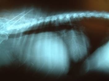
- Thoracic radiographic findings include:
- Space occupying mass with dorsal elevation of trachea and esophagus
- Caudal displacement of cardiac silhouette
- Megaesophagus and aspiration pneumonia with paraneoplastic myasthenia gravis
- Minimal pleural effusion with non-invasive thymoma
- Pleural effusion which may obscure mass with invasive thymoma
- Pulmonary metastasis
- Ultrasonography: mixed echogenicity with cavitation compared to homogenous hypoechogenicity with LSA
- Advanced imaging (i.e., CT or MRI)
- FNA or needle-core biopsy: predominance of lymphocytes rather than epithelial cells may confuse diagnosis
- Immunohistochemistry may be required for definitive diagnosis (cytokeratin)
+ Clinical Staging
- Sub-staging based on presence of paraneoplastic syndromes:
- P 0: no paraneoplastic syndrome
- P 1: myasthenia gravis
- P 2: non-thymic malignant tumor
Treatment
+ Surgery
- Exploratory thoracotomy required to differentiate non-invasive and invasive thymoma
- Median sternotomy usually required due to size of tumor, but lateral intercostal thoracotomy can be used for smaller lesions or in cats (although adjacent rib resection sometimes required)
- Non-invasive thymomas do not adhere to intrathoracic structures and removed using blunt-sharp dissection
- Cranial vena cava and phrenic nerves are located along the craniodorsal aspect of cranial mediastinal mass
- Invasive thymomas usually invade vital structures and are difficult surgical candidates
- Venous grafts are used in humans, and has been reported in the dog, for thymomas invading the cranial vena cava
+ Radiation Therapy
- Thymomas are radiation-sensitive tumors in cats, dogs, and humans
- 75% response rate in cats and dogs with thymomas, including 20% CR
- Lymphoid component of thymoma may determine completeness of response
- Adverse effects: pneumonitis and pericarditis
+ Chemotherapy
- Chemotherapy is usually ineffective, but can be attempted in combination with corticosteroids for invasive thymoma
- Partial and complete responses are uncommon
- Corticosteroids may provide either prolonged stable disease or even partial or complete response
- Response to corticosteroids is due to cytotoxic effects on T lymphocytes which can represent a large non-neoplastic component of thymoma
- Cisplatin, ifosfamide, corticosteroids, doxorubicin, maytansine, cyclophosphamide, vincristine, and procarbazine are used in single or multiple agent protocols in humans with invasive and metastatic thymomas
+ Other Treatment
- Immunosuppressive therapy or anticholinesterase treatment for myasthenia gravis
- Motility drugs, H 2 antagonists, and antibiotics for prophylactic management of megaesophagus
+ Prognosis
- Prognosis is excellent for non-invasive thymoma without megaesophagus as majority of dogs die from unrelated causes and 1-year survival rate 83%
- MST for dogs with radiation therapy: 248 days
- Poor prognostic factors include age, megaesophagus, and histology:
- Age < 8 years associated with a significantly decreased survival time (MST 1 day v 18 days)
- MST for dogs with megaesophagus is 4 days
- MST for lymphocyte-rich thymomas is significantly better than other histologic types of thymoma (MST 237 days v 13 days for differentiated epithelial, 1 day for undifferentiated epithelial, 1 day for oval cells, and 0 days for spindle cells)
- Prognosis is guarded for invasive thymoma or thymoma associated with megaesophagus
- 28% (2/7) local tumor recurrence rate
CRANIAL MEDIASTINAL
THYMOMA
Stages
| I | Well-encapsulated with no microscopic capsular invasion |
| II | Microscopic capsular invasion or gross invasion into adjacent tissue, mediastinal pleura, and fat |
| III | Gross invasion into adjacent organs, pericardium, great vessels, and lungs |
| IVa | Pericardial or pleural dissemination |
| IVb | Distant metastasis through hematogenous or lymphatic routes |