

PATHOPHYSIOLOGY
+ General Considerations
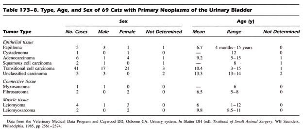
- Epithelial tumors, in particular TCC, are the most common tumors of the urinary bladder and account for up to 92% of feline bladder tumors
- Other urinary bladder tumors include SCC, ADC, rhabdomyosarcoma, FSA, CSA, leiomyosarcoma, HSA, chemodectoma, and benign tumors such as leiomyoma and fibroma
+ Transitional Cell Carcinoma
- Dogs excrete large quantities of tryptophan metabolites in the urine
- However, cats metabolize tryptophan differently and do not excrete urinary tryptophan metabolites
Location
- TCC are either diffuse or located in the fundus or ventral bladder wall in cats
- TCC are most frequently located in the trigonal area in dogs resulting in urinary tract obstruction
DIAGNOSIS
+ Clinical Signs
- Dysuria, hematuria, pollakiuria, and stranguria
- Vaginal discharge
- Urinary obstruction
- Incontinence
- Urinalysis and Urine Sediment Cytology
Urinalysis and Urine Sediment Cytology
+ General Considerations
- Hematuria and proteinuria are consistent findings on urinalysis due to ulceration of the urothelial mucosa
- Bacteruria, pyuria, and positive urine cultures are common in cats
- Bladder wash cytology may improve diagnosis capabilities by reducing contaminants
+ Urine Sediment Cytology
- Neoplastic transitional cells are difficult to differentiate from reactive transitional cells
- Mesenchymal tumors exfoliate poorly and are rarely diagnosed with analysis of urine sediment
+ Blood Tests
- Hematology and serum biochemistry findings are usually normal or non-specific
- Mild to moderate normochromic, normocytic anemia can be caused by either hematuria or bone marrow suppression secondary to chronic disease
- Uremia may result from neoplastic obstruction of urinary outflow or age-related renal failure
Imaging Studies
+ Survey Radiographs
- Magining techniques include survey abdominal and thoracic radiographs, contrast radiography, and CT
- Survey radiographs: sublumbar lymph node enlargement, renomegaly, and metastatic disease in the pulmonary parenchyma or skeleton, particularly lumbar vertebrae and pelvis
- Positive contrast cystography is useful for identification of mucosal abnormalities and space occupying lesions
- Excretory urogram is indicated to determine the location and extent of obstructive urinary tract disease when the urethra cannot be catheterized
+ Ultrasonography
- Recommended to determine the location and extent of bladder involvement, regional lymph node size and appearance, and involvement of adjacent anatomical structures such as the colon
- Superior to excretory urography and double-contrast cystography in detecting TCC
+ Biopsy
- Required for definitive diagnosis of urinary tract tumors
- Techniques include FNA, needle biopsy, catheter biopsy, cystoscopic, and open surgery
- Percutaneous biopsy procedures are not recommended due to the risk of tumor seeding
- Inflammation secondary to necrosis and ulceration is common and may result in false-negative findings
TREATMENT
+ Surgery
- Surgical techniques for management of bladder tumors include:
- Palliative procedures: tube cystostomy ± partial cystectomy
- Curative-intent procedures: partial cystectomy and total cystectomy with urinary diversion
- Cystostomy tube can be placed percutaneously or with either laparoscopic or open surgery
- Complications: stranguria, pollakiuria, hematuria, urine leakage around the stoma, and vesicoureteral reflux which predisposes to ascending UTI and tumor seeding of the upper urinary tract
+ Adjunctive Management
Role of chemotherapy and radiation therapy is unknown