

+ General Considerations
- Primary lung tumors are uncommon in dogs accounting for 1% of all tumors
- Mean age 9.3-11.5 years
- No sex or breed predilection ± higher incidence in Boxer and Beagle
+ Types of Lung Tumors
- Carcinomas account for 97% of primary lung tumor in dogs with ADC more prevalent and SCC uncommon
- Carcinomas are subclassified according to their location (i.e., bronchial, bronchoalveolar, and alveolar)
- Primary lung tumors are graded as well, moderately, and poorly differentiated in dogs
- Degree of differentiation based on scoring system of nuclear pleomorphism, mitosis, and necrosis
- 46% of primary pulmonary carcinomas are grade I, 43% grade II, and 10% of carcinomas are grade III
- Malignant histiocytosis or generalized histiocytic sarcomas often present with solitary or multiple pulmonary masses
- Benign tumors (i.e., chondroma and lipoma) and primary sarcomas are rare
- Lymphomatoid granulomatosis is a very rare tumor
+ Metastasis
- Metastasis can occur via lymphatic, airway, hematogenous, and trans-pleural routes
- 50% metastatic rate with ADC, 90% with anaplastic carcinomas, and 100% with SCC at necropsy in dogs
- Lungs and regional lymph nodes are the most common sites of metastasis, but others include pericardium, heart, pleura, appendicular skeleton, synovium, liver, adrenal gland, pancreas, sternal and axillary lymph nodes, abdominal lymph nodes, kidney, brain, and esophagus
CLINICAL FEATURES
+ Clinical Signs
- Non-productive coughing, exercise intolerance, and other respiratory signs (i.e., dyspnea and tachypnea)
- Systemic signs include lethargy and weight loss
- Peracute presentation for hemothorax, pneumothorax, or malignant pleural effusion is uncommon
+ Paraneoplastic Syndromes
- Hypertrophic osteopathy has been reported in cats and dogs
- Paraneoplastic leukocytosis (which resolved following lung lobectomy) has been reported in 1 dog
- Hypercalcemia of malignancy has been reported in cats and dog
DIAGNOSIS
+ Thoracic Radiographs
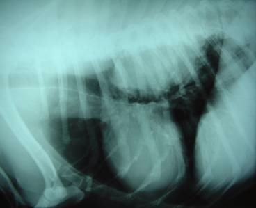
- Lung tumors appear as a well-demarcated and spherical solitary ± cavitary mass in dogs
- Caudal lung lobes are most commonly affected
- Multiple masses may be present in dogs with primary pulmonary LSA, malignant histiocytosis, and lymphomatoid granulomatosis
- Diffuse lesions are present in up to 37% dogs with primary lung tumors
- Uncommon findings: multiple or miliary lesions, hilar lymphadenopathy, and pleural effusion
+ Advanced Imaging
- CT and MRI provide more accurate information on staging for resectability and detection of occult metastasis and hilar lymph node enlargemen
+ Bronchoscopy
- indications: brush cytology of centrally located lesions extending into the bronchus
- trans-tracheal wash and bronchoalveolar lavage can be performed but are usually only diagnostic in diffuse LSA
+ Other Diagnostic Tests
- Thoracocentesis if pleural effusion
- Trans-thoracic FNA for larger lesions with a peripheral location, but larger tumors often have a necrotic centre resulting in a false-negative result
- Trans-thoracic FNA has an 80% accuracy rate, but is associated with 12% mortality rate in cats and dogs
- However, cytologic or histopathologic diagnosis is usually not required as results will not change treatment options (i.e., lung lobectomy)
+ Clinical Staging
37%-55% dogs have T 1 disease at surgery, 22%-25% have lymph node metastasis, and 8% have distant metastasis
Treatment
+ Surgical Management
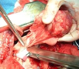
- Lateral thoracotomy (4th-6th intercostal) for small to medium-sized lung tumors and hilar lymph node biopsy
- Median sternotomy for large tumors and inspection of other lung lobes, but lymph node biopsy is more difficult
- Lymph node aspirate or biopsy is recommended as neoplastic infiltration may not be clinically apparent
- Partial lobectomy can be performed for peripheral tumors, but complete lung lobectomy preferred
- Lobectomy can be performed with either stapling equipment or individual ligatures
+ Chemotherapy
- Systemic chemotherapy may offer some benefit
- Intracavitary chemotherapy ± sclerosing agents (i.e., talc or tetracycline) have been used for malignant effusions
- Inhalant chemotherapy using paclitaxel or doxorubicin has been investigated in dogs with primary and metastatic lung tumors:
- Responses in sarcomas but not carcinomas to doxorubicin
- Responses in both sarcomas and carcinomas with paclitaxel
- Response rate 27% (6/22) with 22.5% PR (5/22) and 4.5% CR (1/22)
- Intermittent and non-productive cough for 1-10 days in 50% dogs following inhalant doxorubicin
- Allergic reactions common following paclitaxel administration
PROGNOSIS
+ General Considerations
MST 361 days for dogs
+ Tumor Location
- Dogs with peripheral lung tumors have significantly better survival time than tumors involving entire lung lobe
- Mean ST for peripheral tumors: 16 months
- Mean ST for entire lung lobe tumors: 8 months
+ Tumor Size
- Dogs with small tumors have a significantly longer survival time than large tumors
- Mean ST for tumors < 5 cm diameter: 20 months
- Mean ST for tumors > 5 cm diameter: 8 months
+ Clinical Signs
- Dogs with no clinical signs have a significantly better survival and DFI than dogs with clinical signs
- Asymptomatic tumors: MST 545 days and median DFI 461 days
- Clinical tumors: MST 240 days and median DFI 177 days
+ Clinical Stage or Lymph Node Involvement
- Dogs with lymph node metastases have a significantly worse survival time and median DFI:
- Tumors without lymph node involvement: MST 452 days and median DFI 351 days
- Tumors with lymph node involvement: MST 26 days and median DFI 6 days
+ Tumor Histology
- Dogs with ADC have a better prognosis than SCC as SCC is often diffuse at diagnosis
- MST for ADC: 19 months
- MST for SCC: 8 months
- Low-grade papillary (or bronchoalveolar) carcinoma has a MST of 494 days v 44 days for other cell types
- 50% metastatic rate with ADC, 90% with anaplastic carcinomas, and 100% with SCC at necropsy in dogs
+ Tumor Grade
- Primary lung tumors are graded as well, moderately, and poorly differentiated in dogs
- 46% of primary pulmonary carcinomas are grade I, 43% grade II, and 10% of carcinomas are grade III
- Histologic grading has prognostic significance in dogs:
- Grade I lung tumors: median DFI 493 days and MST 790 days
- Grade II lung tumors: median DFI 191 days and MST 251 days
- Grade III lung tumors: median DFI 0 days and MST 5 days
LUNG TUMORS
| T0 | No evidence of neoplasia | T1 | Solitary lung tumor surrounded by lung or visceral pleura - Primary Tumor | T2 | Multiple lung tumors of various sizes | T3 | Lung tumor invading adjacent tissue | N0 | No evidence of lymph node involvement | N1 | Bronchial lymph node involvement - Node | M0 | No evidence of metastasis | M1 | Evidence of distant metastasis with site specified - Metastasis | N2 | Distant lymph node involvement |