

+ GENERAL CONSIDERATIONS
- Joint tumors are usually malignant
- Joint tumors is a misnomer as tumors of the articular cartilage have not been reported
- Joint is secondarily involved following invasion of subchondral bone at articular margins
+ Tumor Types
- Synovial cell sarcoma is the most common joint tumor
- However, this is controversial as synovial cell sarcoma accounts for only 27% of joint tumors in other studies
- Other reported joint tumors include FSA, rhabdomyosarcoma, myxoma and myxosarcoma, malignant fibrous histiocytoma, liposarcoma, OSA, undifferentiated sarcoma, MCT, SCC, melanoma, and solitary plasmacytoma
+ Survey Radiographs
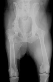
- Joint tumors have a similar radiographic appearance
- Definitive diagnosis requires histopathology ± immunohistochemistry
- Radiographic appearance: partially lobulated soft tissue mass adjacent to the joint, tendon sheath, or bursa
- Bone destruction in 11%-45% (± 72%) dogs with and ≥ 2 bones involved in 100%
- Bone destruction may be:
- Smooth and well-delineated due to pressure necrosis from expansile mass
- Less distinctive lysis due to soft tissue infiltration of bone
- Bone destruction can appear as either permeative lysis or punctate bone loss
- Soft tissue mineralization in 15%-32% in humans, however, not reported in animals with joint tumors
+ Immunohistochemistry
- Histopathologic diagnosis of joint tumors, particularly synovial cell sarcoma, is controversial
- Cytokeratin is used to diagnose synovial cell sarcoma
- Cell morphology and CD18 are used to diagnose histiocytic joint tumors
- Histologic pattern is used to diagnose synovial myxomas
- Actin is used to diagnose malignant fibrous histiocytoma
SYNOVIAL CELL SARCOMA
Pathophysiology
+ General Considerations
- Malignant tumor arising from mesenchymal cells within the tenosynovial tissue of joints, bursa, or tendon sheath
- Stifle is most common site followed by the elbow, shoulder, antebrachiocarpal, talocrural, and hip joints
- Local tumor recurrence common after conservative surgery
- 8%-32% metastatic rate at diagnosis and up to 41% metastatic rate at necropsy
- Metastatic sites: regional lymph node and lungs
- Synovial cell sarcoma is uncommon in cats with the majority of tumors being benign, but 2 cases of metastatic synovial cell sarcomas reported to the regional lymph nodes are lungs
+ Histology
- 2 distinct cell populations: epitheliod or spindle
- histologic subclassification: monophasic (i.e., 1 cell type) and biphasic (both cell types)
- STS, CSA, FSA, malignant fibrous histiocytoma, malignant mesenchymoma, rhabdomyosarcoma, and liposarcoma have similar histologic features with a malignant fibroblastic component
- Histiocytic appearance has also been described
+ Signalment
- Mean age 6-8 years (range, 1-12 years)
- Sex predisposition: ± male with a male-to-female ratio of 1.5:1
- Breed predisposition: Flat-Coated Retriever and Golden Retriever
+ Treatment
- Limb amputation
- Role of chemotherapy is unknown
+ Prognosis
- Median DFI significantly decreased with incomplete resection or conservative surgery (4.5 months v 30 months)
- MST 93 days with no treatment
- MST 455 days to 17 months overall
- Prognostic factors include clinical stage, surgical dose, histologic grade, and positive cytokeratin staining
- Metastatic disease at diagnosis significantly decreases MST (< 6 months v 36-48 months)
- Radical surgery significantly increases MST (840 days v 455 days for marginal resection)
- Grade III synovial cell sarcoma has a significantly worse prognosis (7 months v 48 months for grade I and 36 months for grade II synovial cell sarcomas)
- Mean survival times for other joint tumors are 31.8 months for synovial cell sarcoma, 30.7 months for synovial myxoma, 5.3 months for synovial histiocytic sarcoma, and 3.5 months for other sarcomas
JOINT TUMORS
| T0 | No evidence of neoplasia | T1 | Tumor well defined with no invasion of surrounding tissue - Primary Tumor |
| T2 | Tumor invading soft tissues |
| T3 | Tumor invading bone and joints |
| M0 | No evidence of metastasis |
| M1 | Evidence of distant metastasis with site specified - Metastasis |
| N0 | No evidence of regional lymph node involvement |
| N1 | Regional lymph node involvement - Node |
| N2 | Distant lymph node involvement |